Figure 1.

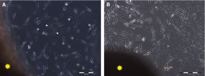
Representative images of fibroblasts in vitro. A, outgrowth from the skin fragment (yellow star) of spindle‐shaped (white asterisks) and star‐shaped (white arrowheads) fibroblasts in culture. B, fibroblasts in culture reached confluence in a time ranging between 14 and 21 d. Scale bar length is 200 µm
