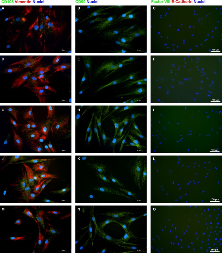
Figure 3.

Representative images of immunocytochemistry for the in vitro expression of mesenchymal (A‐B, D‐E, G‐H, J‐K and M‐N) and epithelial (C, F, I, L and O) markers by dermal fibroblasts from neck (A‐C), breast (D‐F), arm (G‐I), abdomen (J‐L), and thigh (M‐O). Scale bar length is 50 (A‐B, D‐E, G‐H, J‐K and M‐N) or 100 µm (C, F, I, L and O)
