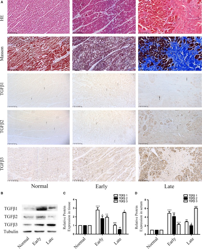
Figure 1.

A, The HE staining showed that the coagulative necrosis of cardiomyocytes indicated the early phase of MI and the appearance of collagens indicated the late phase of MI. In Masson staining, the silk‐like fibres indicated the early phase of MI and the appearance of collagen fibres indicated the late phase of MI. The immunohistochemical staining showed the qualitative analysis of TGFβ1, TGFβ2 and TGFβ3 in the normal and MI tissues (n = 6, each group). B,C, Expression levels of TGFβ1, TGFβ2 and TGFβ3 in human tissue after heart infarction according to Western blot and semiquantitative analysis. The results are shown using tubulin as an endogenous control. The data are presented as the mean ± SEM (n = 6, each group). *P < 0.05 versus the normal group; **P < 0.01 versus the normal group; ***P < 0.005 versus the normal group; #P < 0.05 versus the early group; ##P < 0.01 versus the early group; ###P < 0.005 versus the early group. D, TGFβ1, TGFβ2 and TGFβ3 expression levels in human serum after myocardial infarction according to ELISA. The data are presented as the mean ± SEM (n = 6, each group). *P < 0.05 versus the normal group;**P < 0.01 versus the normal group; ***P < 0.005 versus the normal group; #P < 0.05 versus the early group; ##P < 0.01 versus the early group; ###P < 0.005 versus the early group
