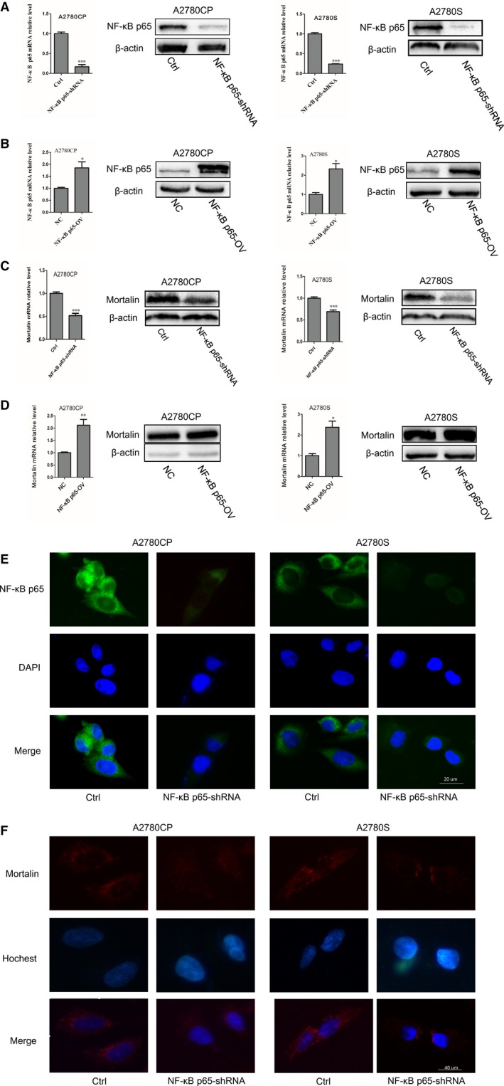
Figure 2.

NF‐κB p65 regulates the expression of mortalin. (A) NF‐κB p65 mRNA and protein expression were assessed by real‐time quantitative PCR and Western blotting in NF‐κB p65‐shRNA cells. (B) NF‐κB p65 mRNA and protein expression were assessed in NF‐κB p65 overexpression cells. (C) Mortalin mRNA and protein expression were assessed in NF‐κB p65‐shRNA cells. (D) Mortalin mRNA and protein expression were assessed in NF‐κB p65 overexpression cells. (E) NF‐κB p65 expression (green) was assessed by immunofluorescence with anti‐NF‐κB p65 primary antibody. DAPI was used to stain nucleus (blue). (F) Mortalin expression (red) was assessed by immunofluorescence with anti‐mortalin primary antibody. Hochest was used to stain nucleus (blue). *P < 0.05, **P < 0.01, ***P < 0.001
