Figure 1.

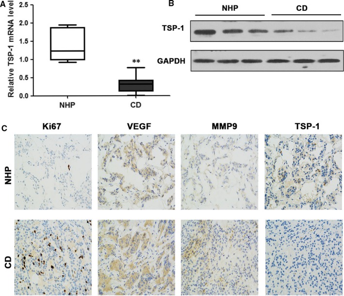
Relative TSP‐1 expression in pituitary corticotrophs. A, The expression of TSP‐1 mRNA in Cushing's disease (CD, n = 33) and normal human pituitary (NHP, n = 7) tissue. B, Western blotting of TSP‐1 protein levels in normal pituitary and CD tissue samples. C, Immunohistochemical staining for Ki67, VEGF, MMP9 and TSP‐1 in representative normal pituitary and corticotroph adenoma (magnification, ×200). Representations of at least three biological replicates are presented (mean ± SEM; **P < 0.01)
