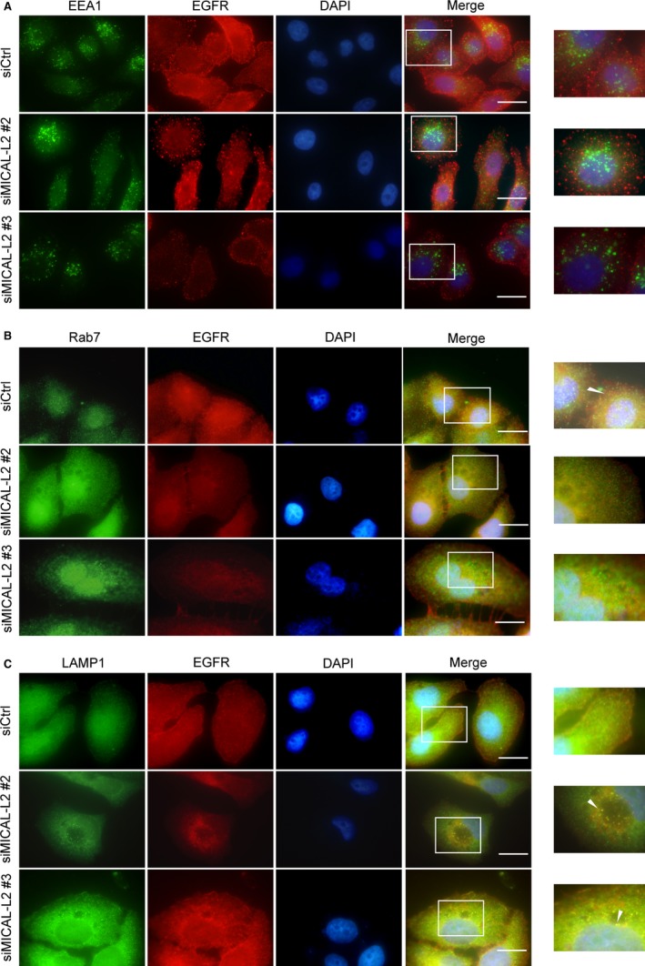
Figure 4.

Effect of MICAL‐L2 on EGFR cellular localization. After transfected with control siRNA or siMICAL‐2, SGC‐7901 cells were immunostained by antibodies against EEA1 (A), Rab7 (B) or LAMP1 (C). All endocytic markers are shown in green. EGFR is shown in red. Nuclei (blue) were visualized by DAPI. The yellow colour indicated the colocalization. Scale bar, 5 μm
