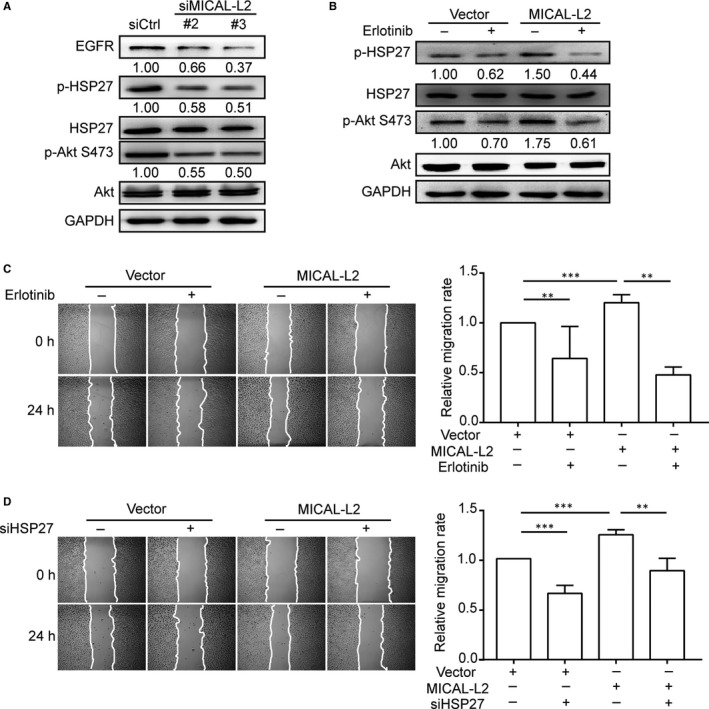
Figure 5.

Effect of MICAL‐L2 on EGFR/HSP27 signalling pathways. A, BGC‐823 cells transfected with control siRNA or siMICAL‐L2 were in serum‐free media overnight and protein levels of EGFR, p‐Akt, p‐HSP27 were detected by Western blotting. B, Cells transfected with empty vectors or MICAL‐L2 plasmids were incubated with 1 μmol/L Erlotinib for 24 h. Then proteins extracted from the lysates were subjected to Western blotting to detect the expression of p‐Akt, p‐HSP27. C, SGC‐7901 cells overexpressing MICAL‐L2 were pre‐treated with 1 μmol/L Erlotinib for 24 h, then migration activity of the cells was analysed. D, SGC‐7901 cells overexpressing MICAL‐L2 were pre‐treated with siHSP27, migration activity was analysed. **P < 0.01, ***P < 0.001
