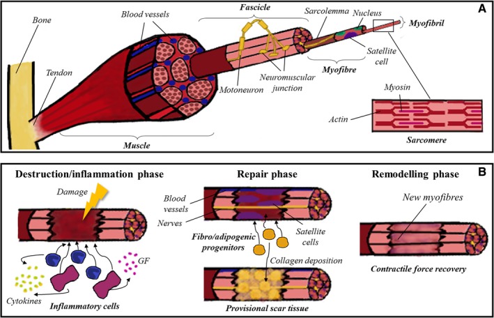
Figure 1.

Skeletal muscle tissue. A, Skeletal muscle tissue comprises several bundles of aligned myofibres, each one containing thousands of myofibrils. In the image it is detailed the ultrastructure of the sarcomere, which is the final responsible of the contraction of the muscle due to the movement of myosin and actin myofilaments. B, The repair process of skeletal muscle is divided into three overlapping phases: destruction/inflammation, repair and remodelling. In the first stage, inflammatory cells are recruited to the damaged zone, secreting cytokines and growth factors which attract satellite cells. Then, adjacent blood vessels and nerves invade the healing area, and fibro/adipogenic progenitors co‐operate to form the provisional scar tissue. Finally, new myofibres are formed, and the repairing process concludes when contractile force is recovered
