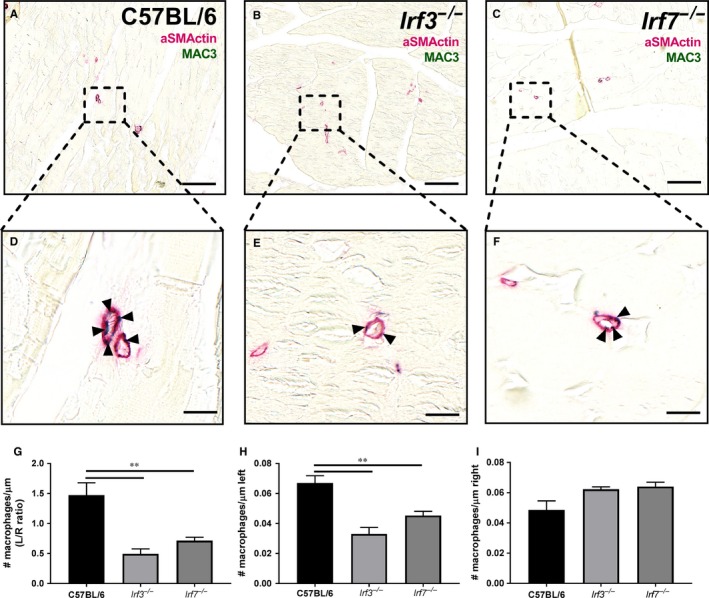
Figure 4.

Macrophages around arterioles. An immunohistochemical double staining was performed on adductor muscles of Irf3−/−, Irf7−/− and C57BL/6 mice sacrificed 28 days after HLI. aSMActin (pink) was used to show the arterioles and MAC3 (green) to show the macrophages around the collaterals. Representative images of the left ischemic adductor muscle of (A) C57BL/6 mice, (B) Irf3−/− mice and (C) Irf7−/− mice stained with aSMActin and MAC3 is shown (scalebar = 100 μm) and a zoom in of the arterioles of the concerned (D) C57BL/6 mice, (E) Irf3−/− mice, (F) Irf7−/− mice adductor muscle is shown (scalebar = 10 µm). Arrows indicate were the macrophages are located in the arterioles. (G) Ratio (left ischemic/right non‐ischemic) of the number of macrophages per μm circumference of arterioles in the adductor muscle is shown. (H) Number of macrophages per μm circumference of arterioles in the left ischemic adductor muscle is shown (I) Number of macrophages per μm circumference of arterioles in the right non‐ischemic adductor muscle is shown. Data is presented as mean SEM; **P < 0.01, Mann‐Whitney test was used. C57BL/6 n = 5, Irf3−/− n = 7, Irf7−/− n = 7
