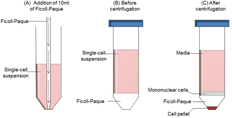
Figure 1: Separation of mononuclear cells using Ficoll-Paque media and density gradient centrifugation.

(A) 10ml of Ficoll-Paque is added to 30ml of single-cell suspension using a 10ml serological pipet and pipet aid. Slow and gentle release (indicated by the dashed arrows) of the Ficoll-Paque will result in a distinct separation between the two layers. (B) Before the density gradient centrifugation, the layer of single-cell suspension is above the Ficoll-Paque media. (C) After centrifugation, TILs and monocytes are found in the interface between the media and Ficoll-Paque, while the pellet consists of tumor cells and the erythrocytes.
