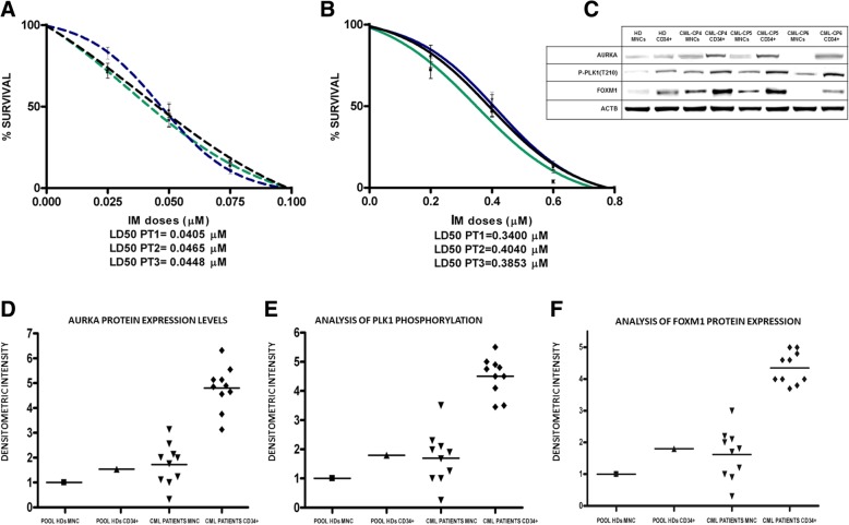
Fig. 4.
Aurora A/PLK1/FOXM1 axis is hyper-activated in CD34+ compartment from CML patients at diagnosis compared to a pool of 8 HD. a-MNCs from three CML patients at diagnosis were sensitive to IM administration with LD50 ranging from 0.0405 to 0.0465 μM; b- Ph1+/CD34+ cells separated from the same three patients exhibited a relative resistance to IM, with LD50 ranging from 0.3400 to 0.4040 μM; c-d-e-f-Aurora A overexpression, PLK-1 hyper-activitation and FOXM1 overexpression is restricted to CD34+ compartment of ten CML patients. Notably, Aurora A and FOXM1 protein expression and PLK1 hyper-phosphorylation were significantly higher in CD34+ cells compared to MCF of HD (panels c, d, e and f), suggesting Aurora A, PLK1-FOXM1 axis is a stemness component in the hematopoietic tissue. In panels d, e and f the values of protein expression and phosphorylation in MNCs and CD34+ cells of individual CML patients relative to the HD pool were obtained by comparison of band densitometry analysis (see Materials and Methods section for details)

