Fig. 5.
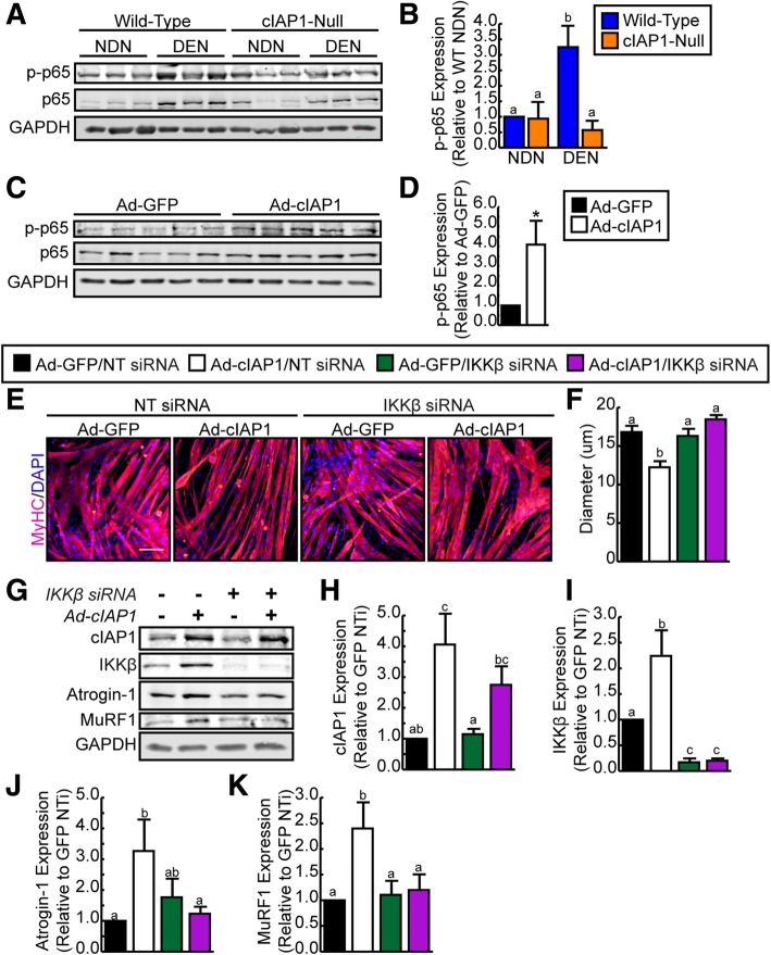
cIAP1 induces atrophy through upregulation of classical NF-κB signaling. a Representative Western blots of phospho-p65, p65, and GAPDH expression in gastrocnemius muscle from non-denervated (NDN) and denervated (DEN) hind limbs of C57BL/6 and cIAP1-null mice. b Quantification of phospho-p65 expression normalized with total protein and relative to wild-type non-denervated (WT NDN) muscle (n = 3). c Representative Western blots of phospho-p65, p65, and GAPDH expression in C2C12 myotubes infected with adenovirus expressing GFP (Ad-GFP) or human cIAP1 (Ad-cIAP1) for 24 h. d Quantification of phospho-p65 expression normalized with total protein and relative to Ad-GFP infected myotubes (n = 5). e Nascent (DM day 2) C2C12 myotubes were transfected with non-targeting (NT) siRNA or siRNA targeting murine IKKβ, allowed to form mature myotubes (DM day 4) and then infected with Ad-GFP or Ad-cIAP1 for 24 h. Myotubes were stained with MyHC (pink) and nuclei were counterstained with DAPI (blue). Representative images are shown. Scale bar = 50 um. f Myotube diameter of myotubes stained as in e (n = 5). g Representative Western blots of cIAP1, IKKβ, Atrogin-1, MuRF1, and GAPDH expression in cells cultured as described in e. Quantification of h cIAP1, i IKKβ, j Atrogin-1, and k MuRF1 protein expression normalized with total protein and relative to NT siRNA transfected/Ad-GFP infected C2C12 myotubes (n = 5). Data is the mean ± SEM, *p < 0.05, means with no common letters are significantly different from each other (p < 0.05)

