Fig. 12.

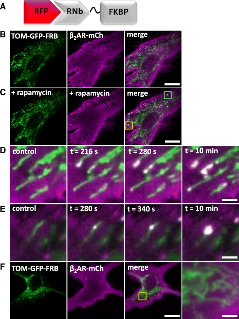
Recruitment of proteins to native PM-mitochondria MCS using RNb-FKBP. a Schematic of RNb-FKBP fusion bound to RFP. b, c HeLa cells co-expressing RNb-FKBP, mitochondrial TOM70-GFP-FRB and β2AR-mCh were imaged using TIRFM before (b) and after (c) treatment with rapamycin (100 nM, 10 min). Scale bar 10 μm. d, e Enlarged images from C of the yellow box (d) and cyan box (e) show punctate recruitment of β2AR-mCh to individual mitochondria at the indicated times after addition of rapamycin. Scale bars 1.25 μm. f TIRFM images of HeLa cells co-expressing mitochondrial TOM70-GFP-FRB and β2AR-mCh in the presence of rapamycin (100 nM, 10 min) show no recruitment in the absence of co-expressed RNb-FKBP. The yellow box shows a region enlarged in the subsequent image. Scale bars 10 μm (main images) and 2.5 μm (enlargement). Results (b–f) are representative of 5 independent experiments
