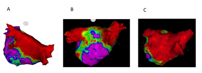
Figure 2. A shows left atrial posterior wall voltage mapping with dense homogeneous isolation of the pulmonary veins and posterior wall. B shows voltage mapping (0.05-0.5mV) following surgical ablation (pre-endocardial). The bilateral pulmonary veins are isolated and posterior wall is partially isolated with gaps in floor line. C is a different hybrid ablation patient also pre-endocardial ablation; as compared to middle panel posterior wall is densely isolated. All voltage maps 0.05-0.5mV.