. 2018 Apr 26;40(1):44–52. doi: 10.1590/2175-8239-JBN-3838

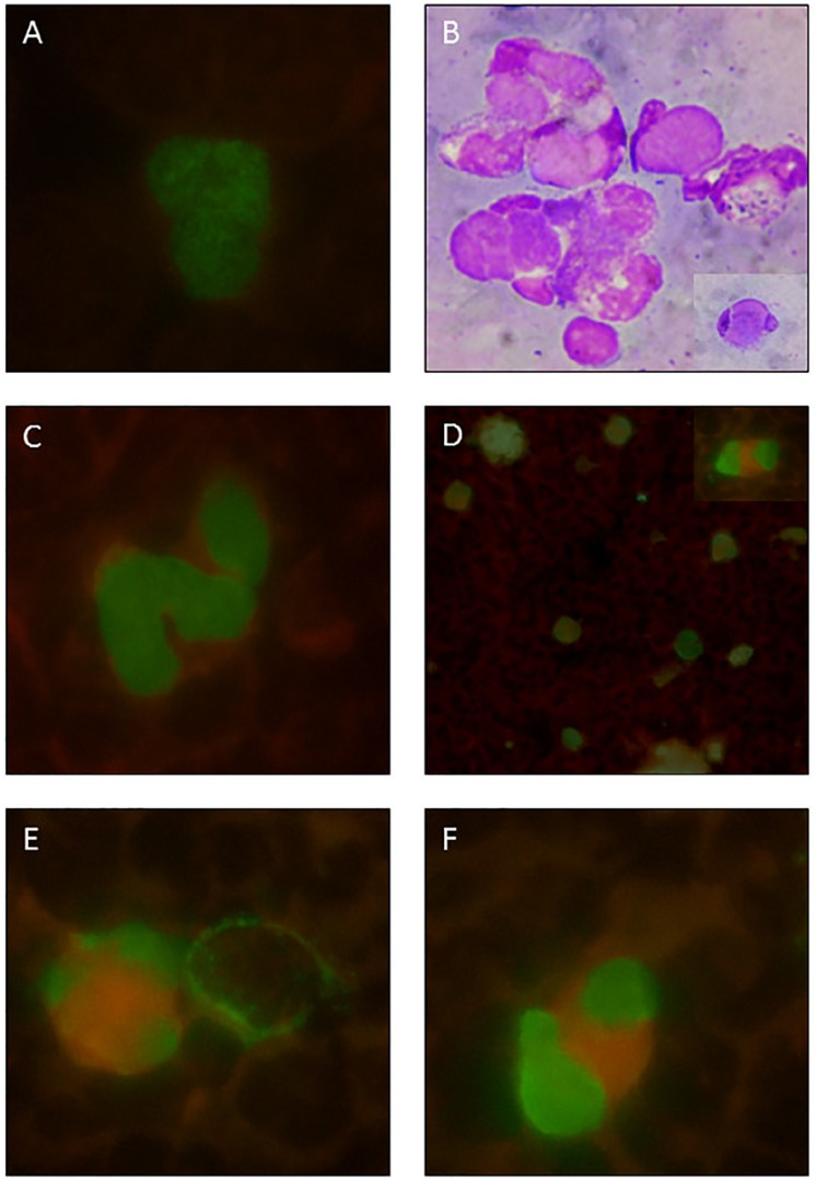
Figure 1. Representative photomicrographs of positive pp65 antigenemia (immunofluorescence). A) pp65 positive neutrophil from a renal transplant patient infected with CMV, used as a reference due to its typical nuclear granular appearance. B) LE cells: neutrophils containing cytoplasmic amorphous inclusions and peripheral nucleus (Wright staining); a detailed view is shown at the lower right corner. C) From case 5: pp65 positive neutrophils with a smooth granular appearance. D) From case 6: panoramic view of pp65 positive neutrophils and cells similar to LE cells; a detailed view is shown at upper right corner. E) and F) From the same SLE case 6: similar dysmorphic aspects of vacuolated cells, not usually seen in transplanted kidney patients.

Figure 1