Figure 1.
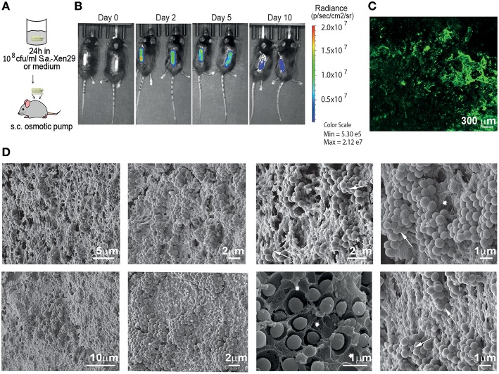
Mouse model for implant-associated infection. (A) Experimental setup. (B) Biofilm implant-associated infection caused by S. aureus Xen29 in C57Bl/6 mice was monitored in real time for 10 days. Images were taken every 24–48 h. Mice were anesthetized with isoflurane during imaging procedures. Luminescent counts were collected for an exposure time of 30 s within each measured region of interest (ROI) and quantified with Living Image software. (C) Representative Light Sheet micrograph from the surface of immunostained osmotic pumps post-explantation, showing anti-S aureus antibody in green. (D) Images recorded by scanning electron microscopy of colonized implant surfaces. Excavations with the shape of bacterial cells could be found in the matrix (asterisks) as well as bacterial cells with altered geometry (arrows). These are representative images for six mice under the same experimental conditions.

