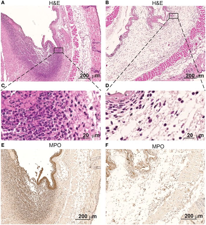
Figure 2.
Skin infiltrates increase when in contact with S aureus Xen29. Microscopic evaluation at different magnification of skin samples on day 2 after implantation of osmotic pumps colonized (A,C,E) or non-colonized with S aureus Xen29 (B,D,F). H&E staining (A–D) and anti-MPO (E,F) immunostaining of tissue surrounding S. aureus colonized pumps. These images are representative for 6 mice per experimental condition.

