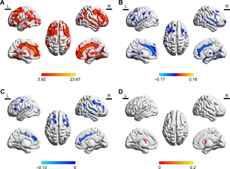
Figure 1.
Differences in GMD.
Notes: (A) GMD in WML patients. (B) GMD in cortical regions of WML-VaD patients. (C) GMD in cortical regions of WML-MCI. (D) GMD in caudate of WML-MCI patients.
Abbreviations: GM, gray matter; GMD, gray matter density; WML, white matter lesion; VaD, vascular dementia; MCI, mild cognitive improvement.

