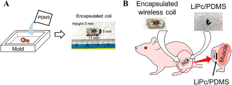
FIGURE 5.

Preparation for in vivo EPR oximetry. A, The wireless coil is placed in a mold that is bigger than the required size. Then the PDMS solution is poured in the mold, keeping the wireless coil in the center. After a day, the mold was removed and the PDMS sides are cut and chamfered to the required dimension and shown in the right side. B, The LiPc/PDMS were tied to the encapsulated wireless coil using surgical suture. This was placed in the lateral side of the left hind leg by making a 5-mm incision in the subcutaneous area
