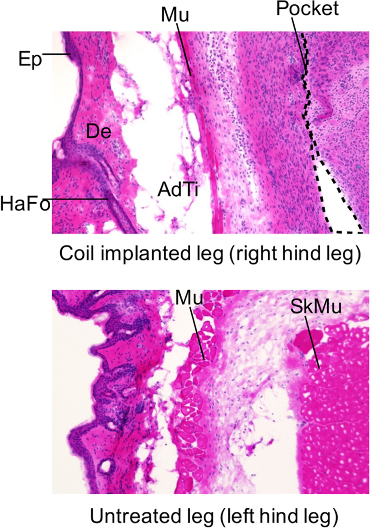
FIGURE 9.

Histological sections of tissue surrounding the wireless coil and that of tissue of untreated leg. Each tissue was excised from the mouse 26 days after the wireless coil was implanted. Each abbreviation in these images indicates the region as follows: AdTi, adipose tissue; De, dermis; Ep, epidermis; HaFo, hair follicle; Mu, panniculus carnosus muscle; Pocket, wireless coil pocket; SkMu, skeletal muscle
