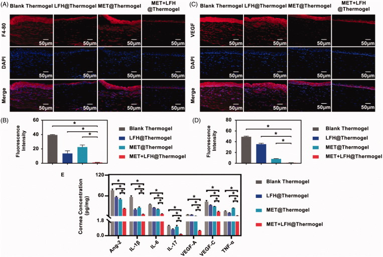
Figure 6.
Anti-angiogenic assessment of different treatments on seventh day after alkali burn. (A) Immunofluorescence staining of the cornea with a specific macrophage maker-F4/80 antibody (red color) and (B) the quantitative analysis (scale bar = 50 μm). The results are presented as mean ± SD (n = 6). *p < .05. (C) Immunofluorescence staining of VEGF (red color) in the cornea and (D) the quantitative analysis (scale bar = 50 μm). The results are presented as mean ± SD (n = 6). *p < .05. (E) Concentrations of Ang-2, IL-1β, IL-6, IL-17, VEGF-A, VEGF-C, and TNF-α in the corneas. The results are presented as mean ± SD (n = 6). *p < .05.

