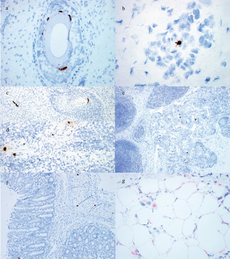
Figure 4.
Detection of PCV3 by ISH in weaned pig cases. (a) PCV3 in hair follicle epithelium of the 3-week-old pig from Case no. 2. (b) PCV3 in an endothelial cell of a small arteriole in the dermis of the 3-week-old pig from Case no. 2. (c) PCV3 in cardiac myocytes and smooth muscle of an arteriole surrounded by inflammation in the 25-day-old pig from Case no. 7. (d) PCV3 in smooth muscle of an arteriole surrounded by inflammation in a 6-week-old pig from Case no. 8. (e) PCV3 in smooth muscle of multiple arterioles surrounded by inflammation and gut-associated lymphoid tissue of the 25-day-old pig from Case no. 7. PCV3 in the (f) smooth muscle and lamina propria of the large intestine and (g) adipocytes of the panniculus of the 3-week-old pig from Case no. 2.

