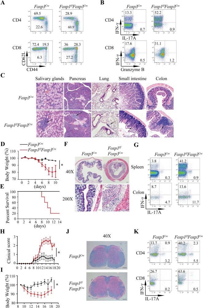
Fig 4. Foxp1 regulates Treg suppressive function.
(A) Flow cytometry analysis of CD44 and CD62L expression in conventional CD4+ T cells (YFP−CD4+) and CD8+ T cells in the spleens of 28-week-old Foxp3Cre and Foxp1f/fFoxp3Cre mice; numbers adjacent to the outlined areas represent the percentages of gated cells. (B) Intracellular staining of cytokines in conventional CD4+ and CD8+ T cells in the spleens of 28-week-old Foxp3Cre and Foxp1f/fFoxp3Cre mice; numbers adjacent to the outlined areas represent the percentages of gated cells. (C) Hematoxylin and eosin staining of salivary gland, pancreas, lung, small intestine, and colon sections. The magnification is ×100. Black arrows indicate the areas of immune cell infiltration. Scale bars, 100 μm. (D-G) Foxp3Cre and Foxp1f/fFoxp3Cre mice (n = 6) were treated with 2.5% DSS, and the severity of colitis in mice was evaluated by loss of body weight (D), survival curve at indicated time points (E), the representative hematoxylin and eosin staining of colon sections (×40 and ×200, respectively; scale bars, 200 μm and 50 μm, respectively) (F), and the intracellular staining of cytokines in CD4+ T cells in the spleens and the colons (G). (H-K) Sex- and age-matched Foxp3Cre and Foxp1f/fFoxp3Cre mice (n = 9–10) were induced with EAE by immunization with MOG peptide and pertussis toxin, and the severity of EAE was evaluated by the clinical score (H), loss of body weight (I), Luxol fast blue-hematoxylin and eosin staining of spinal cord sections (×40, scale bars, 200 μm) (J), and the intracellular staining of cytokines in T cells from the spinal cords and the brains (K). Data in (A-C, H-K) represent at least three independent experiments. Data in (D-G) represent two independent experiments. Data in (D, H, I) are mean ± SEM, *P < 0.05 (two-tailed Student t test). Data associated with this figure can be found in the supplemental data file (S1 Data). CD, cluster of differentiation; DSS, dextran sulfate sodium; EAE, experimental autoimmune encephalomyelitis; Foxp1, forkhead box P1; IFNγ, interferon gamma; IL, interleukin; MOG, myelin oligodendrocyte glycoprotein; Treg, regulatory T; YFP, yellow fluorescent protein.

