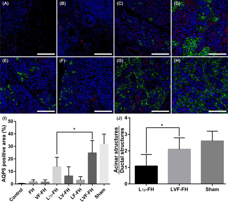
Fig. 5. LVF-FH promoted aquaporin expression throughout the newly formed glandular tissue and increased the acinar to ductal structure ratio similar to unwounded tissues.
Immunostaining for AQP5 (green, acinar marker) and K7 (red, ductal marker) in wounded SMG treated without scaffold (A), or with FH (B), VF-FH (C), L1p-FH (D), LV-FH (E), LF-FH (F) and LVF-FH (G). Unwounded glands served as sham controls (H). Specimens were analyzed using a confocal Zeiss LSM 700 microscope at 40× magnification (bars = 200 μm). Positive area of AQP5 (I) and the ratio of acinar and ductal structures (J) were calculated on day 20 post-surgery using ImageJ and analyzed using one-way ANOVA (*p < 0.01, n = 12) and Dunnett’s post-hoc test for multiple comparisons to the L1p-treated group.

