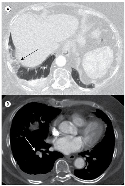
Figure 2. In A, computed tomography angiography image, with lung window settings, showing a lesion characteristic of pulmonary infarction with the reversed halo sign (black arrow) in the subpleural region of the right lower lobe, comprising heterogeneous areas of low attenuation. A small pleural effusion is also seen on this side. In B, computed tomography angiography image, with mediastinal window settings, showing a small filling defect (white arrow) at the emergence of the segmental branch that connects the right pulmonary artery to the lateral basal segment of the lower lobe.

