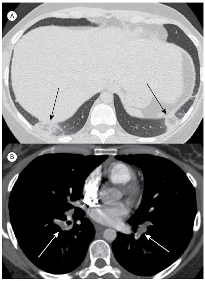
Figure 3. In A, computed tomography angiography image, with lung window settings, showing lesions characteristic of pulmonary infarction with the reversed halo sign (black arrows) in the subpleural region of the lower lobes, containing reticulation; the lesion in the right lower lobe is oval, and the one in the left lower lobe is round. In B, computed tomography angiography image, with mediastinal window settings, showing filling defects (white arrows) at the emergence of the pulmonary arteries.

