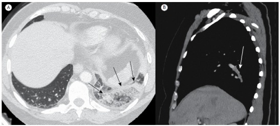
Figure 5. In A, computed tomography angiography image, with lung window settings, showing oval lesions characteristic of pulmonary infarction with the reversed halo sign (black arrows) in the subpleural region of the left lower lobe, comprising heterogeneous areas of low attenuation. In B, sagittal reconstruction, with mediastinal window settings, showing an extensive filling defect (white arrow) in the segmental branch that connects the left pulmonary artery to the posterior basal segment of the lower lobe.

