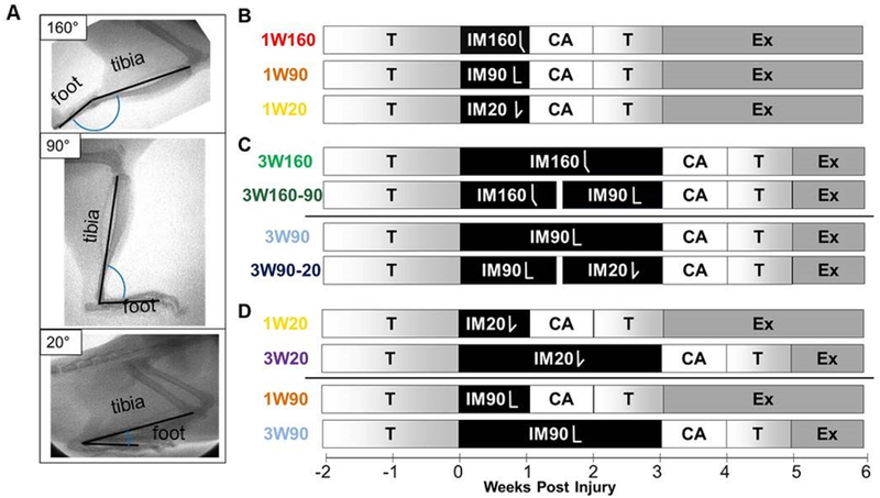
Figure 1. Study Design.

(A)Ankle immobilization angle was confirmed with fluoroscopy. Following exercise training (T), animals had their central right plantaris removed and right Achilles tendon bluntly transected. After surgery, animals were immediately placed in one of three casts: full plantarflexion (160° tibiopedal angle, IM160), mid-point (90° tibiopedal angle, IM90), and anatomic sitting position (20° tibiopedal angle, IM20). Within each angle, animals were assigned to either one week of immobilization (1W) or three weeks of immobilization (3W). Two groups assigned to 3W underwent manipulation on the 11th day of their 3 week immobilization period (160° to 90°, IM160–90; 90° to 20°, 90–20). After cast removal, animals were allowed cage activity (CA) for one week, followed by one week of treadmill training (T), followed by treadmill running (exercise, Ex) daily until sacrifice. All animals were sacrificed six weeks post-injury (n=16/group). Comparisons were made to compare (B) the effects of increased dorsiflexion during immobilization for 1 week to the standard plantarflexed position (1W160 vs. [1W90 or 1W20]), data shown in Figures 2 and 3; (C) the effects of manipulation of ankle angle during immobilization starting from 160 degrees (3W160 vs 3W160–90) or 90 degrees (3W90 vs 3W90–20), data shown in Figures 4 and 5; and (D) the effects of dorsiflexion immobilization duration at 90 degrees (1W90 vs 3W90) or 20 degrees (1W20 vs 3W20), data shown in Figures 6 and 7.
