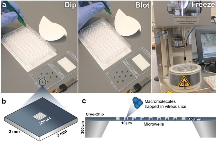
Figure 2.
The preparation of frozen-hydrated macromolecules using the “cryo-EM-on-a-chip” technique. a) Microchips are dipped into the sample containing the macromolecule of interest before the excess aqueous buffer solution is wicked away using Whatman filter paper. Chips are plunge frozen into liquid ethane and used for downstream imaging and analysis. b) Schematic diagram of a Cryo-Chip with integrated microwells. The 2 mm × 2 mm chip has a central imaging region of 500 μm × 500 μm and can be engineered with a variety of microwells designs. c) Side view schematic representation of a Cryo-Chip. The height of the chip is ≈300 μm, while each individual well has a depth of 150 μm and a pitch of 10 μm. The microwells aid in the encapsulation of macromolecules in vitreous ice.

