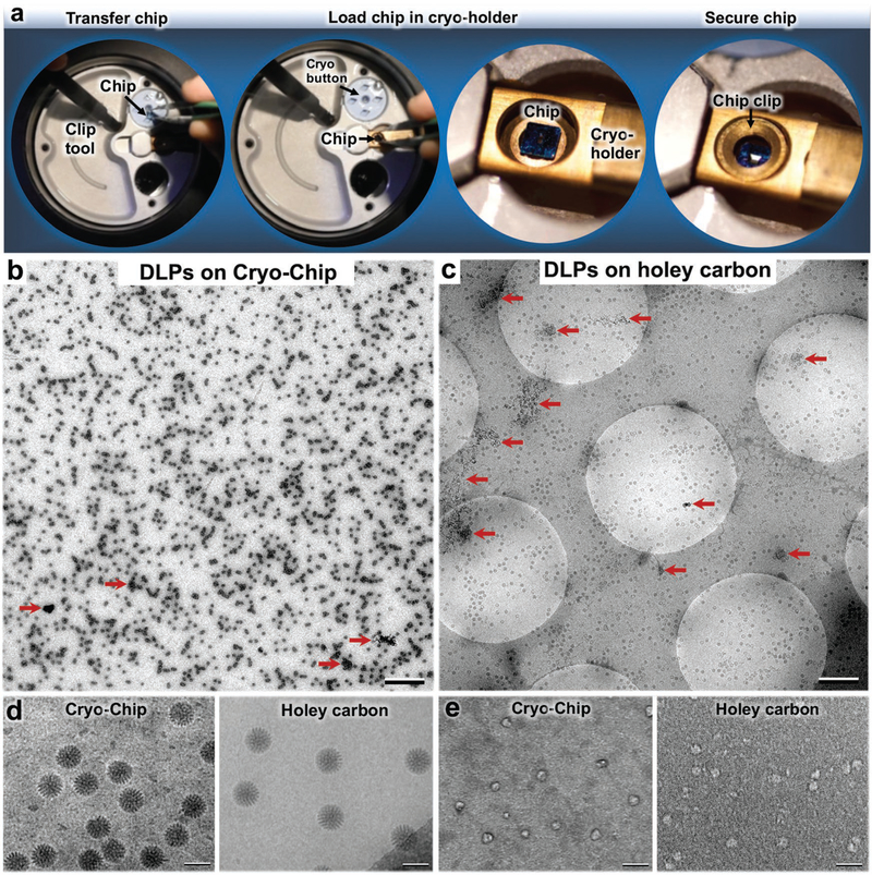
Figure 3.
Cryo-Chip specimen transfer and image comparisons. a) Using a clip tool, Cryo-Chips are transferred from cryogenic storage buttons to the tip of a EM specimen holder, then secured in place using a modified clip ring. A comparison of DLPs prepared on b) Cryo-Chips and c) holey carbon grids revealed a consistent ice layer and a greater number of usable particles on Cryo-Chips. AWIPs are indicated by red arrows. Scale bar is 0.5 μm in panels (b) and (c). d) Samples prepared using SiN substrates demonstrated higher visual contrast for active DLPs prepared on holey carbon films, as previously demonstrated.[19] e) Enhanced image contrast was noted for Cryo-Chips specimens of smaller molecules such as BRCA1-associated protein assemblies. Scale bar is 100 nm.

