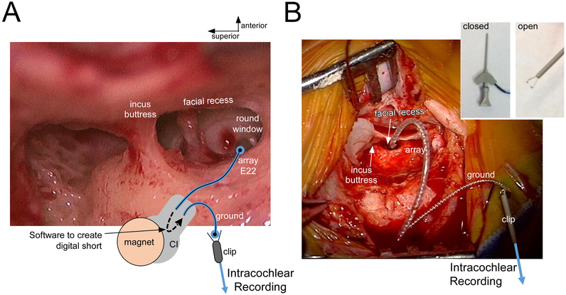
Figure 1.
Intraoperative setup for recording intracochlear ECochG with a Cochlear Corporation array. (A) View through surgical microscope with superimposed schematic diagram. The array’s deepest contact (E22) is digitally shorted to the ground rod contact (ECE1) when the processor is connected and software delivered through the telemetry is used. With this connection made, a clip electrode is connected to the ground rod and fed to the BioLogic recording device. (B) Photograph of the intraoperative recording setup, after the array was fully inserted. The ground and clip are floating above the surgical field (isolated electrically from the patient). Inset: Open vs Closed positioning of the clip electrode.

