Figure 4.
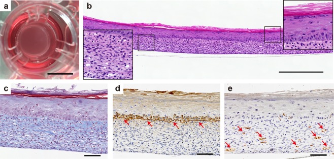
In vitro evaluation of the therapeutic potential of pre-vascularized 3D skin substitutes. (a–e) HUVEC (0.2 × 10⁵ cells/insert) mixed with FN-G-coated NHDF were cocultured, subsequently covered with HEKn, then cultured for up to an additional 7 days. (a) Macroscopic view of the construct in the culture insert. Scale bar: 10 mm. (b–e) Histological and immunohistochemical staining with hematoxylin and eosin (b), Masson’s trichrome (c), anti-laminin 5 (d, arrows = basement membrane), and anti-CD31 (e, arrows = CD31+ blood vessel). Scale bars: 500 μm (b), 100 μm (c–e).

