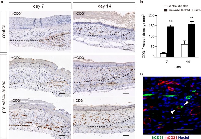
Figure 6.
Quantification of wound vasculature in vivo. (a) Immunohistochemical detection of mouse-specific CD31 (mCD31) and human-specific CD31 (hCD31) at 7 and 14 days after grafting. Dashed lines indicate the skin substitute–host interface. Scale bars: 100 μm. (b) Quantification of CD31+ blood vessels in the dermal areas of grafts. The data are the mean ± SD (n = 5). **p < 0.01 compared with the non-vascularized control (unpaired Student’s t-test). (c) Immunofluorescence image stained for mouse-CD31 (red) and human-CD31 (green) of pre-vascularized substitute-treated wounds at day 14 after grafting. Nuclei were stained with DAPI (blue). White arrowheads indicate human–mouse chimeric vessel. Scale bars: 50 μm.

