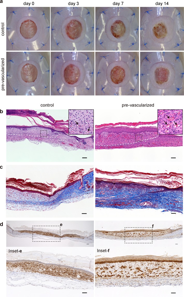
Figure 7.
Pre-vascularization promoted wound healing and remodeling. (a) Macroscopic observation of transplanted control and pre-vascularized substitutes on the backs of immunodeficient mice up to 14 days post-transplantation. (b–f) Histological and immunohistochemical staining with hematoxylin and eosin (b), Masson’s trichrome (c), and HLA-ABC (d–f) of the wounds from the control and pre-vascularized substitutes at 14 days post-transplant. White dashed lines indicate the skin substitute–host interface. Black arrows indicate vessels perfused by mouse blood. Scale bars: 100 μm.

