Fig. 5.
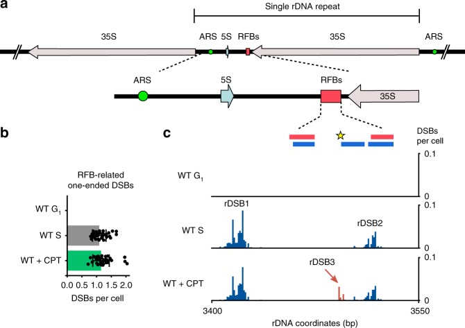
DNA double-strand breaks at replication fork barriers. a Scheme of replication fork barriers (RFBs) at yeast ribosomal DNA locus. b The total number of RFB-related one-ended DSBs (peaks as defined in c calculated from the difference of Watson and Crick strand reads (Methods)). DSBs per cell values and SD are shown (n = 39, Methods). Source data are provided as a Source Data file. c Quantified DSB signal in RFB region. RFB1 and RFB2 are indicated by the red boxes on the top. The blue boxes mark Fob1 protein binding sites mapped in vitro. The yellow star indicates Top1 cleavage site. The signal originating from rDSB-1 and rDSB-2 is presented in blue, the signal from rDSB-3 we discovered is presented in red

