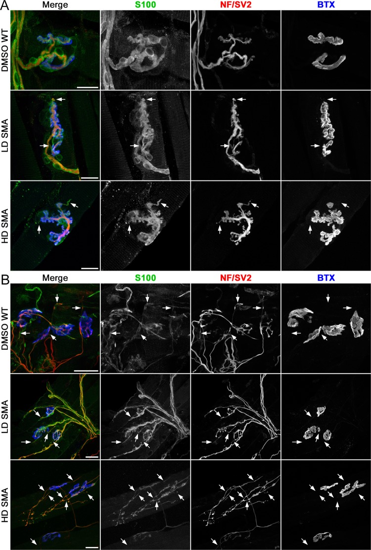
Figure 2.
Terminal sprouting at endplates in contralateral and reinnervated SOL muscles. Confocal maximum projections of endplates after whole mount staining for Schwann cells (S100), axons and nerve terminals (NF/SV2) and AChRs (BTX). Arrows point to terminal sprouts. (A) Contralateral SOL: An example of a NMJ without terminal sprouts in DMSO WT SOL, and two examples of NMJs with two short terminal sprouts each in LD and HD SMA SOL. (B) Denervated SOL: Endplates with more profuse terminal sprouting 7 days after crush denervation in all treatment groups. Scale bars: 20 μm (A). 30 μm (B).

