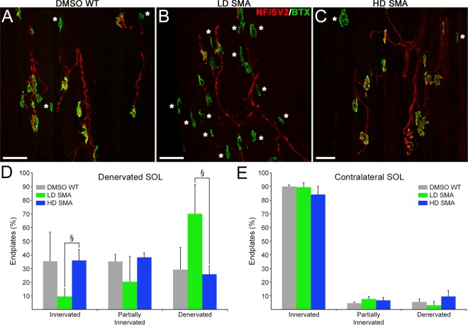
Figure 5.
Slower reinnervation in LD SMA SOL was rescued in HD SMA SOL. (A–C) Representative fields from DMSO WT, LD SMA and HD SMA SOL longitudinal sections stained for AChR (green) and axons/nerve terminals (red), 7d after crush of the tibial branch of the sciatic nerve (Fig. 1). Denervated endplates are indicated by asterisks. Scale bar: 50 μm. (D) Quantification of innervation status on the denervated SOL. Sample sizes: DMSO WT: 2 mice, 144 endplates; LD SMA: 4 mice, 302 endplates; HD SMA: 4 mice, 355 endplates. §p < 0.05, ANOVA with Bonferroni correction. (E) Quantification of innervation status on the contralateral SOL. Sample sizes: DMSO WT: 2 mice, 179 endplates; LD SMA: 4 mice, 312 endplates; HD SMA: 3 mice, 153 endplates. No significant differences were found in innervation status of contralateral muscles between treatment groups.

