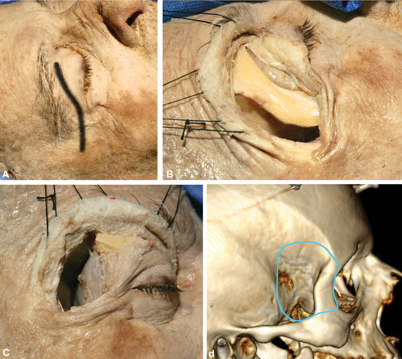
Fig. 1.

Macroscopic part of the approach. ( A ) The skin incision is through the eyelid, starting from the midpupillary line and extending 1 cm behind the lateral orbital ridge, along the zygomatic arch. ( B ) Exposure of the lateral orbital rim in strict subperiosteal fashion. The lateral orbital rim is removed by drilling of the sphenoid ridge and exposure of the frontal, temporal dura and periorbita, followed by two osteotomies, using an oscillating saw, just above the frontozygomatic suture superiorly and at the level of the zygomatic arch. ( C ) Macroscopic view of the surgical field after the osteotomies and after the bone removal extending to the pterion is completed. The frontal, temporal lobes, as well as the periorbital, are visualized. ( D ) Three-dimensional reconstruction using the OsiriX software (version 5.8.1, Pixmeo, Bernex, Switzerland), presenting the limits of the craniotomy (the visualized screws are the markers for the neuronavigation).
