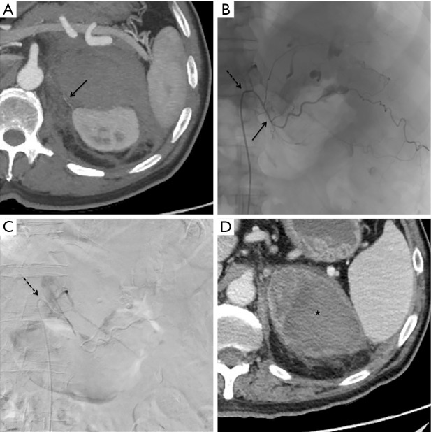
Figure 2.
Patient 1, same patient of Figure 1. Axial maximum intensity projection post-processing CT reconstruction (A): the superior adrenal artery is appreciable (black arrow). Procedural fluoroscopy (B) showing a 5 Fr Cobra catheter (dotted black arrow) at the origin of the left phrenic artery and a 2.7 Fr microcatheter (continuous black arrow) at the origin of superior adrenal artery: similar to CT (Figure 1), multiple foci of contrast agent extravasation are evident and so 300–500 micron microparticles were adopted as embolizing agent. Post embolization digital subtraction angiography (C) after contrast injection from the 5 Fr Cobra catheter (dotted black arrow), revealing no more extravasation in the embolized area. CT scan of control in arterial phase 24 hours after the embolization procedure (D) detecting left adrenal hematoma (black asterisk) without signs of active bleeding.

