
Fig. 1.
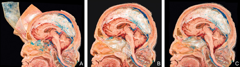
Cadaveric dissections of extracranial pericranial flap (sagittal view). ( A ) Elevated flap with osteotomy at nasion. ( B ) Flap is passed through the bony window below the frontal sinus and along the roof of the ethmoidectomy defect. Note the large dimensions of the flap. ( C ) The flap is placed over the clival defect.