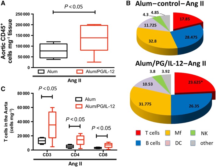
Figure 2.

Characterization of aortic leukocytes following P. gingivalis immunization and low‐dose Ang II infusion. Mice were immunized with an i.p. injection of P. gingivalis formulated with alum and IL‐12 (Alum/PG/IL‐12) and are compared with control mice that received an i.p. injection of alum only (Alum). Following immunization, both groups received a 14 day infusion of a low dose of Ang II infusion (0.25 mg·kg−1·day−1). Flow cytometry was used to quantify total leukocytes (A, CD45+), *P < 0.05 compared to control, n = 8 in each group. Within the CD45+ cells, the percentage of T cells, B cells, macrophages, NK cells and dendritic cells were quantified (B). T cells – CD3+; lymphocytes B (B cells) – CD19+; macrophages (Mf) – I‐Ab+/CD11b+; dendritic cells (DC) – I‐Ab+/CD11c+; and NK cells (NK) – NK1.1+, *P < 0.05 versus control, n = 8 (C). T‐cell infiltration in aortas of P. gingivalis‐immunized mice and controls. T‐cell subpopulations (CD4+ and CD8+ T cells were studied) by flow cytometry following a 14 day infusion of a low‐dose of Ang II, *P < 0.05 compared to control, n = 8 in each group.
