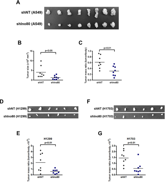
Figure 3. Ino80 is required for tumor formation in mouse xenografts.
A549, H1299, and H1703 cells were infected with NT or Ino80-shRNA viruses and injected subcutaneously into immunocompromised mice. Tumors were dissected 5 weeks after the injection. (A, D, F) Representative image showing the size of the tumors. (B-C) Analysis of tumor size (B) and mass (C, E, G). Data was plotted as Mean ± SEM, and n = 8 in each group. p-value was calculated by two-tailed Student’s t-test.

