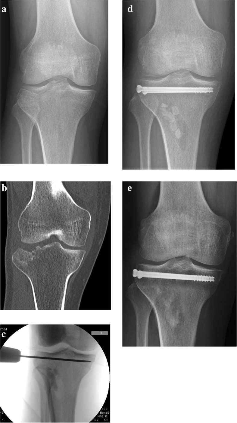
Fig. 1.

a Pre-operative anteroposterior x-rays of a patient with a split compression tibial plateau fracture (Schatzker type III). b Coronal section of computerized tomography scan. c Intra-operative fluoroscopy showing temporary fixation with a K-wire and metaphyseal bone defect filled with bone substitute grafting. d Post-operative anteroposterior x-rays of two cannulated screws fixation. e Postoperative 12 months’ anteroposterior x-rays showing good radiological results and initial resorption of the bone graft
