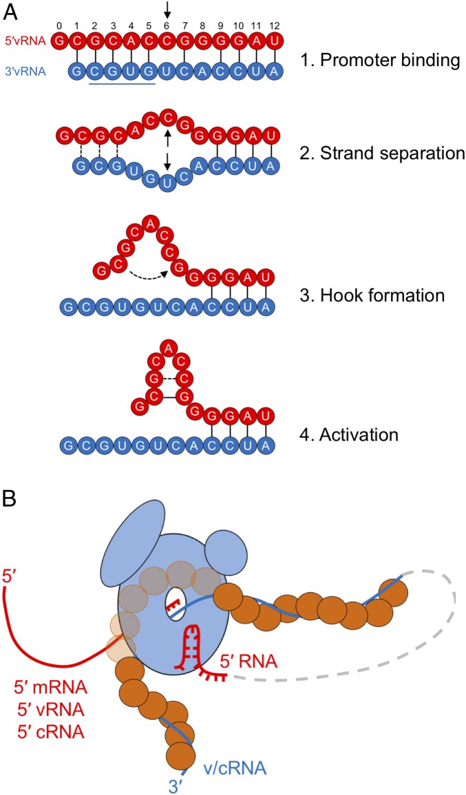
Fig. 5.
Model of arenavirus L activation by a 5′ hook structure present in the terminal genomic (vRNA) and antigenomic (cRNA) RNA segments. (A) Proposed four-step mechanism of L initiation and activation by the 5′ vRNA ligand (red). The underlined 3′ vRNA sequence represents the previously identified motif for promoter recognition by MACV L (20). (B) A model of the activated arenavirus RNA synthesis complex. L (light blue) is shown bound to the 3′ v/cRNA (blue line), which is encapsidated by the viral nucleoprotein (orange circles). The nascent 5′ RNA (red line) is shown exiting to the left of L. The activating 5′ hook (red line) is also illustrated as being bound by L. The intermediate RNP is depicted as a dashed gray line connecting the 3′ RNA and activating 5′ hook functioning in cis.

