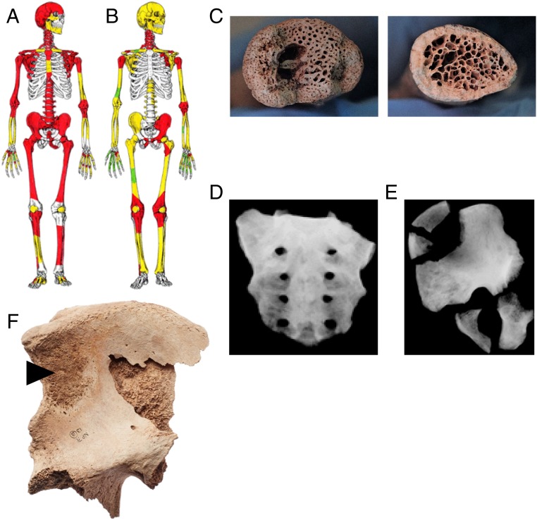
Fig. 1.
Skeletal distribution of macroscopic changes (red), internal lytic changes identified by radiographic analysis (yellow), and unaffected bones (green) of a PDB-like disorder in SK101 (A) and SK29 (B). (C) Macroscopic observation of internal structural changes in the right clavicle of SK37 (Left) compared with the normal cortical and trabecular structure of an unaffected right clavicle (Right; SK50). Radiographic imaging of SK37 sacrum (D) and hip (E). (F) Macroscopic observation of osteosarcoma (arrowhead) in the pelvis of SK29. The extracortical portion of the tumor exhibits a slight radiant alignment of bone that is often referred to as a “sunburst” appearance. Reprinted with permission of the Norton Priory Museum Trust.

