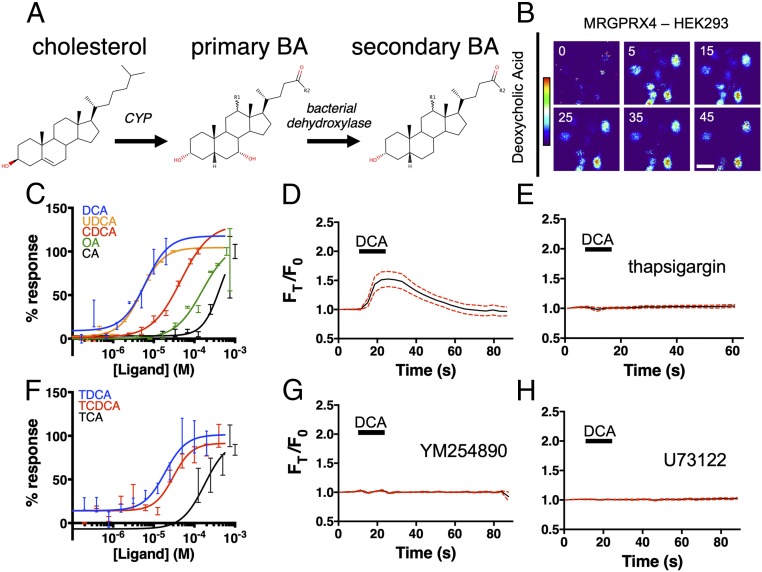
Fig. 1.
BAs activate MRGPRX4, a human sensory neuron-expressed GPCR. (A) The molecular stereostructures of primary and secondary BAs and their metabolic precursor, cholesterol. Major structural differences are highlighted in red. R1 denotes the potential presence of an α-hydroxyl substituent in some BA derivatives. R2 denotes additional substituents that vary among BAs. (B–H) Ca2+ imaging of HEK293 cells stably expressing MRGPRX4. (B) Representative Fura-2 fluorescence heat map images of HEK cells showing changes in intracellular [Ca2+] induced by DCA (10 µM). (Scale bar, 20 µm.) (C and F) Concentration–Ca2+ response curves of BAs against MRGPRX4. Data are a representative experiment of three independent replicates performed in triplicate, depicted as mean ± SEM (C) Concentration–Ca2+ response curves of unconjugated BAs. OA, oleic acid. (F) Concentration–Ca2+ response curves of tauro-conjugated BAs. (D, E, G, and H) Ca2+ imaging traces. A total of 10 µM DCA was added where indicated by black bars. Mean ± 95% CI depicted. n = 15. (E, G, and H) Cells were preincubated with either 10 µM sarco/endoplasmic reticulum Ca2+-ATPase inhibitor thapsigargin (E), 10 µM of the Gαq inhibitor YM254890 (G), or 10 µM PLC inhibitor U73122 (H) for 30 min before imaging.

