
Figure 3.
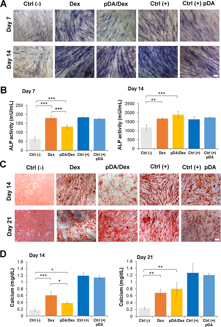
Differentiation of hASCs cultured on gels coated with pDA and treated with dexamethasone. (A) Alkaline phosphatase staining of hASCs seeded on the different gels at day 7 and day 14. A total of five groups were tested. The Ctrl (–) group indicates hASCs cultured on gel without any treatment in culture medium without any osteogenic factor. The Dex group represents hASCs cultured on gel soaked with Dex 0.1 mM in non-osteogenic medium. The pDA/Dex group indicates hASCs cultured on gel precoated with a layer of pDA and soaked afterwards with Dex 0.1 mM. Cells were cultured in the same condition as the Dex group. The Ctrl (+) group represents hASCs cultured on gel without any treatment in osteogenic medium. The Ctrl (+) pDA group displays hASCs cultured on gel precoated with a layer of pDA and cultured in osteogenic medium. Scale bar: 100 μm. (B) ALP quantification at days 7 and 14 for the different groups (n = 5). (C) Alizarin red staining of calcium deposited by hASCs seeded on the different groups at days 14 and 21. Scale bar: 100 μm. (D) Quantification of calcium in the different samples after differentiation of hASCs for 14 and 21 days. (n = 5). The results are reported as a mean plus or minus standard deviation. (n = 5). Single asterisks indicate p < 0.05, double asterisks indicate p < 0.01, and triple asterisks indicate p < 0.001.