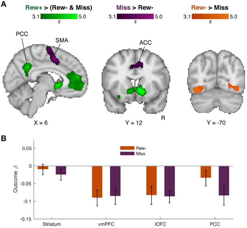
Figure 3: Whole-brain trial outcome contrasts.

(A) Results of whole-brain contrasts for Rew+ trials > Rew− and Miss trials (green), Miss trials > Rew− trials (purple), and Rew− trials > Miss trials (orange). In the reward contrast (green), four significant clusters were observed, in bilateral striatum, ventromedial prefrontal cortex (vmPFC), left orbital-frontal cortex (OFC), and posterior cingulate cortex (PCC). For the Miss trial/motor error contrast (purple), three significant clusters were observed, one single cluster spanning bilateral premotor cortex, supplementary motor area (SMA), and the anterior division of the cingulate (ACC), as well as two distinct clusters in both the left and right inferior parietal lobule (not shown). The Rew− trials > Miss trials contrast showed a single cluster spanning visual cortex. (B) Beta weights extracted from each reward-contrast ROI for the (orthogonal) Rew− and Miss trial outcomes. Error bars = 1 s.e.m. See also Table S1 and Figures S3 and S4.
