
Figure 1. BM-derived MSCs remained viable after coating with polymer.
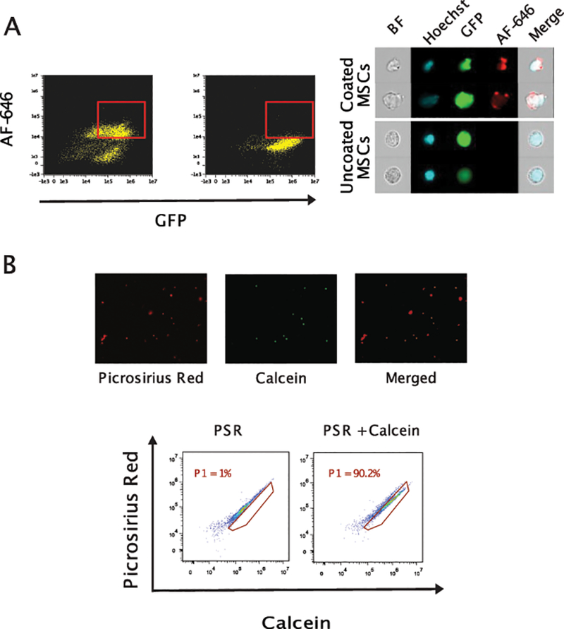
Coated BM-MSCs were visualized by imaging flow cytometer-ImageStream System. Coated and uncoated BM-MSCs were fixed, stained with antibodies against collagen I antibody (AF-647) for the presence of cell surface gelatin coating, and acquired on an ImageStream flow cytometer. (Panel A) Representative FACS plots and images demonstrating nucleated coated GFP+ BM-MSCs with Hoechst33342 and Alexa-Fluor 647 positive staining. (Panel B) Coated cells remained alive after coating process as stained double positively with picosirius red (red for gelatin coating) and calcein (green for viability) as assessed by microscopy (upper panel) and flow cytometry (lower panel).