
Figure 3. Cell coating began to degrade in vitro within 24 hr.
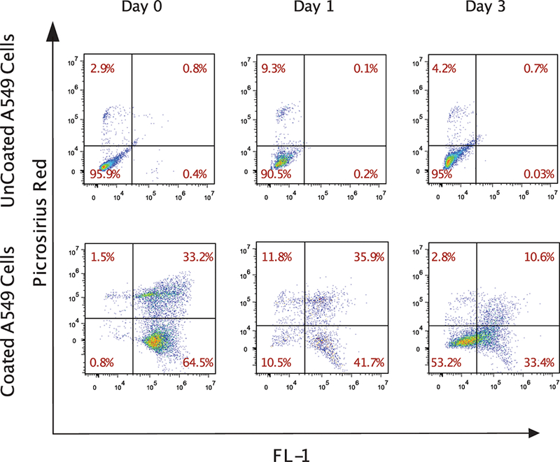
Coated and uncoated control A549 lung carcinoma cells were cultured for up to three days. Coated cells were identified with Picosirius red. The fluorescence in the FL-1 channel of the coated cells was due to the presence of eosin, which was used to initiate polymerization. The plots demonstrate the progressive loss of coating on cultured cells over time.